

Sleep spO2 vs ODI: Troubleshooting Low Oxygen Readings

What you’re likely seeing when sleep spO2 and ODI don’t match
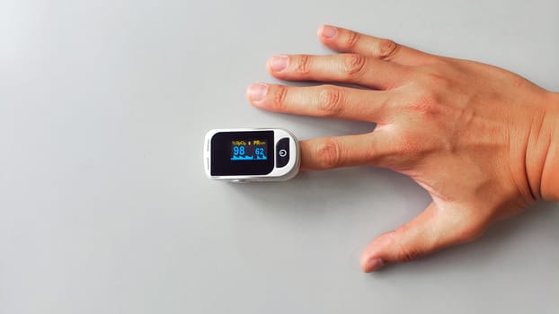
If you’re reviewing your sleep data and noticing that your sleep spO2 looks “okay” while your ODI is high (or the reverse), it can be confusing—and sometimes concerning. Many people start troubleshooting only after they notice symptoms like:
- Waking with dry mouth or headaches, even though average oxygen saturation seems normal
- Frequent awakenings, restless sleep, or feeling unrefreshed
- High ODI events reported as oxygen drops, but your spO2 graph doesn’t show obvious deep dips
- Frequent low spO2 readings that don’t seem to correlate with how you feel
- Inconsistent results across nights (one night looks “fine,” the next looks “bad”)
In real-world use, the mismatch often means one of two things: either your breathing pattern is causing brief desaturations that ODI captures more clearly than average spO2, or your sensor/recording setup is producing artifacts that distort one metric more than the other.
This troubleshooting guide focuses on the practical steps you can take to determine which situation you’re dealing with, then move from simplest fixes to more advanced ones.
How sleep spO2 and ODI differ (and why that matters for troubleshooting)
Before you change anything, it helps to anchor what the metrics actually represent.
Sleep spO2 is oxygen saturation measured continuously (or near-continuously) by a pulse oximeter. Many reports show an “average,” a “minimum,” and a curve across the night.
ODI (oxygen desaturation index) counts how many times your oxygen saturation drops by a defined amount within a defined time window—commonly 3% or 4% drops. The key point: ODI is sensitive to brief desaturation events, even if your overall average spO2 stays relatively stable.
So if ODI is high but average spO2 is normal, you may be experiencing many short desaturations—often linked to obstructive sleep apnea or unstable breathing. If spO2 is low but ODI is modest, you may be dealing with slower, sustained drops, measurement drift, or signal quality issues.
Either way, troubleshooting starts with measurement accuracy and then moves to breathing-related causes.
Most likely causes of “sleep spO2 vs ODI” confusion
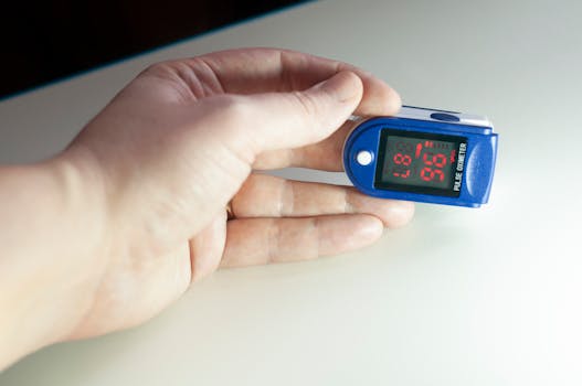
In practice, mismatches usually come from one (or more) of the following categories.
1) Sensor positioning, fit, and signal quality problems
Pulse oximeters are sensitive to motion, poor perfusion, cold hands/feet, and loose placement. If the device loses a clean signal for even short periods, the software may estimate values that don’t reflect your true oxygenation.
Common triggers include:
- Finger probe placed too loosely or on a moving limb
- Using a wrist or clip sensor when the signal is marginal
- Cold fingers causing weak blood flow
- Sleep positions that compress the sensor (bending the wrist, leaning on the hand)
- Dry skin or nail polish interfering with optical readings
- Low battery or inconsistent device contact
2) Breathing events that cause brief desaturation (ODI spikes)
If your ODI is high, one of the most common physiological explanations is obstructive sleep apnea (OSA) or related breathing instability. OSA often produces repeated cycles of airway collapse, oxygen dips, and then partial recovery—precisely the pattern ODI is designed to count.
Symptoms that strengthen this cause include loud snoring, witnessed apneas, frequent awakenings, and daytime sleepiness.
3) Artifact from device settings or reporting thresholds
Some systems allow different desaturation thresholds (e.g., ODI3 vs ODI4). If you compare reports from different devices or different firmware/software versions, the ODI number may not be directly comparable.
Even within the same brand, the device may apply different smoothing or event detection rules. That doesn’t mean anything is “wrong,” but it can make nights look inconsistent.
4) Sustained oxygen issues that affect spO2 more than ODI
Less commonly, you might see low spO2 with a relatively lower ODI if oxygenation is reduced more continuously rather than in repeated sharp drops. This can relate to lung or circulation conditions, high altitude, certain medications, or other medical causes.
If you have known respiratory disease, oxygen issues deserve careful attention and professional guidance.
Step-by-step troubleshooting and repair process
Use this sequence. The goal is to determine whether the mismatch is primarily a measurement problem, a breathing pattern problem, or both.
Step 1: Confirm you’re looking at the same night and the same metric definitions
Start with the boring but essential checks.
- Make sure the spO2 and ODI are from the same recording session.
- Check whether your ODI is based on a 3% or 4% desaturation threshold (ODI3 vs ODI4). If the app doesn’t label it, look for settings or device documentation.
- If you’re comparing two devices, don’t assume ODI numbers are directly interchangeable.
Practical example: You may see ODI “high” after switching from a finger clip to a wrist sensor. The wrist sensor can be less stable during side-sleeping, and the threshold algorithm may count motion-related dips as desaturations.
Step 2: Evaluate signal quality before you interpret oxygen
Most sleep devices provide some indication of signal quality, pulse strength, or a “good/poor” indicator. If you have access to raw data or quality flags, use them.
Even without raw data, you can infer signal problems if:
- spO2 repeatedly jumps up and down unrealistically
- the pulse waveform (if shown) is erratic
- the lowest spO2 values occur during periods of movement
- the pulse rate and oxygen curve look out of sync
If you suspect signal problems, fix them first. Don’t chase physiology with a bad measurement.
Step 3: Fix sensor placement and contact (do this for 2–3 nights)
Make targeted adjustments. Don’t change everything at once—change one variable, then repeat.
- Finger clip sensors: Place the probe on a finger with good circulation. Remove nail polish if relevant. Keep the finger warm before bed (even 5–10 minutes can help). Ensure the clip is snug but not painful.
- Wrist sensors: Keep the wrist level with your heart when you lie down. Avoid sleeping with the wrist bent sharply under your body.
- Timing: Put the sensor on at least 10–15 minutes before you fall asleep so the device can stabilize.
- Movement reduction: If you toss and turn, consider a tighter bedding setup or a sleep position that reduces compression on the sensor.
- Skin and temperature: If your hands are cold, warm them. Cold perfusion is a common cause of poor oximetry signals.
Track whether the spO2 curve becomes smoother and whether ODI events cluster at specific times (e.g., early night vs later night).
Step 4: Run a “daytime sanity check” for the device
Before relying on overnight data, check whether the device behaves plausibly when you’re awake and still.
- Measure while seated quietly for 2–3 minutes.
- Note whether spO2 stabilizes within a short time and whether readings look consistent.
- Move slightly and see how quickly the device recovers. If it takes a long time or produces erratic values, the sensor may not be reliable for sleep.
This step doesn’t diagnose physiology. It diagnoses whether the measurement is trustworthy.
Step 5: Reduce known confounders for 1–2 nights
Some factors can change oxygenation and breathing patterns, and they can also affect sensor contact.
- Avoid alcohol within 4–6 hours of bedtime. It can worsen airway collapse and desaturation events.
- Be consistent with sleep position. If side-sleeping improves oxygen data, you may see ODI drop when you maintain that position.
- If you use a humidifier, keep it consistent so airway dryness doesn’t change your breathing stability.
- Don’t compare nights with major illness (colds, congestion) to nights without symptoms.
Solutions from simplest fixes to more advanced fixes
Once you’ve done the core troubleshooting steps, apply fixes in order. Stop when your data stabilizes and your symptoms improve.
Simple fixes: improve measurement reliability
- Warm your extremities: Cold hands are one of the fastest ways to degrade oximeter signal quality. Aim for stable readings before sleep.
- Re-seat the sensor: If you see frequent “spikes” or abrupt drops, remove and reapply. Use the most stable placement your device supports.
- Check for compression: If you sleep on your hand or bend your wrist, reposition to avoid pressure on the sensor.
- Remove nail polish: For finger probes, this can matter. If you can, use a clean nail surface for testing.
- Use the same device settings: Keep ODI threshold settings consistent (ODI3 vs ODI4) and don’t mix different report types.
Intermediate fixes: reduce breathing-related desaturation triggers
If the measurement looks stable (smooth spO2 curve, believable pulse waveform, consistent readings across nights), then address likely breathing contributors.
- Positional therapy (start with simple changes): If you notice ODI events occur mostly when you’re on your back, try training side-sleeping. Even a basic positional strategy—like an arrangement that makes back-sleeping uncomfortable—can reduce events for many people.
- Optimize nasal airflow: If you’re congested, treat it consistently (e.g., saline rinse before bed) rather than inconsistently. Nasal resistance can worsen airway collapse in susceptible people.
- Sleep schedule consistency: Irregular sleep can increase fragmentation and worsen breathing stability. Keep bedtime within a 60–90 minute window for 1–2 weeks while you monitor trends.
- Weight and metabolic factors (slow-burn): If you’ve had recent weight gain, it can increase OSA risk over time. You won’t fix this in a week, but trends matter.
Practical example: You run a 3-night sensor placement test and see ODI remains high only when you sleep on your back. After positional changes for 7 nights, your ODI drops by a meaningful amount and your morning headaches reduce—even though average spO2 was similar each night. That pattern points toward event-driven desaturation rather than a persistently low baseline.
Advanced fixes: validate the physiology with better diagnostics
If ODI remains high after you’ve ruled out sensor artifacts, you need higher-confidence evaluation. Home pulse oximetry can be a useful signal, but it can’t identify the cause of desaturation by itself.
Consider these next steps:
- Request a formal sleep evaluation: If you suspect OSA (high ODI, snoring, witnessed pauses, daytime sleepiness), ask for a sleep study. This is the definitive way to connect desaturation events with respiratory effort and airflow.
- Use an oximeter with reliable signal quality: If your current device frequently reports poor signal or you see obvious artifacts, switching to a device designed for overnight oximetry can improve confidence. Focus on signal quality indicators and proven sensor stability rather than raw marketing claims.
- Check for respiratory contributors: If you have lung disease, neuromuscular conditions, or symptoms like shortness of breath, a clinician may evaluate oxygenation with additional testing.
- Medication review: Sedatives and certain medications can worsen airway tone. If your desaturation worsens after a medication change, discuss it with a prescribing clinician.
Advanced troubleshooting is about moving from “data mismatch” to a clinically meaningful explanation.
When replacement or professional help is necessary
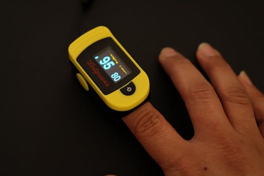
Use these thresholds as decision points. If you’re unsure, err toward professional assessment—especially when symptoms are present.
Replace or stop using the current sensor when you see persistent measurement problems
It’s reasonable to replace the device or stop relying on its oxygen metrics if:
- Your readings are highly inconsistent across multiple nights despite consistent setup
- You repeatedly see implausible drops (e.g., sharp dips that quickly rebound without matching your breathing or pulse)
- The device frequently reports poor signal quality
- Daytime sanity checks show unstable or erratic values when you’re still
In that case, the mismatch may be a measurement artifact rather than a real oxygen issue.
Seek professional help urgently if you have concerning symptoms
Get prompt medical advice if you have any of the following alongside oxygen concerns:
- Chest pain, severe shortness of breath, or fainting
- New or worsening neurologic symptoms (confusion, severe headache)
- Signs of significant oxygen impairment during the day
- Known cardiopulmonary disease and new desaturation patterns
Even if you’re only seeing it on sleep data, don’t ignore these red flags.
Schedule a sleep evaluation if ODI stays high after measurement fixes
If you’ve improved sensor placement for 2–3 nights and the pattern persists, professional evaluation is the next rational step—especially if you have symptoms like snoring, witnessed apneas, or daytime sleepiness.
High ODI with consistent event patterns is often linked to sleep-disordered breathing that can be treated. The key is confirming the cause with appropriate testing rather than guessing from a single oxygen metric.
Putting it all together: a targeted troubleshooting workflow you can follow
Here’s a practical workflow that mirrors how many clinicians think about oximetry data:
- Verify definitions and consistency: Confirm the ODI threshold and that you’re comparing the same kind of report.
- Check signal quality: Look for erratic spO2 behavior or poor pulse/signal indicators.
- Correct the measurement: Improve placement, warmth, and avoid compression for 2–3 nights.
- Confirm stability: Run a short daytime sanity check for reliability.
- Interpret the pattern: If ODI events persist with stable measurement, treat it as likely breathing-related.
- Escalate appropriately: If it persists, arrange a sleep evaluation or medical assessment based on symptoms and risk factors.
That approach prevents you from chasing your tail between spO2 and ODI numbers when the real issue is either sensor integrity or a treatable breathing disorder.
How to monitor progress without overreacting to single-night noise
Oxygen metrics can vary night to night. Your goal isn’t to “win” one night—it’s to see a trend after you fix setup variables.
For most people, a sensible monitoring plan is:
- Do 2–3 nights of consistent sensor setup and note whether spO2 curves become smoother and ODI events stabilize.
- Then apply one change at a time (like positional therapy or nasal airflow support) for 7–14 nights while tracking whether ODI decreases and symptoms improve.
- If you’re making medical changes or starting treatment, follow a clinician’s guidance and use objective data to confirm improvement.
If the mismatch between sleep spO2 and ODI reverses repeatedly without any setup changes, the measurement is likely unstable. If the mismatch is consistent and tied to symptoms or position, it’s more likely physiological.
Common real-world scenarios and what they usually indicate

Scenario 1: ODI is high, average spO2 is normal, and you snore
This pattern often points toward repeated brief desaturation events related to obstructive sleep apnea. If your sensor setup is stable and ODI stays elevated across nights, professional sleep evaluation is usually the most direct next step.
Scenario 2: spO2 shows repeated low dips, but ODI is low
That can happen when oxygenation is drifting lower without meeting the desaturation threshold counts, or when the device is producing artifacts (motion, poor contact, cold). Start by improving sensor contact and checking signal quality. If the pattern persists, get medical advice—especially if you have lung disease or symptoms.
Scenario 3: One night looks dramatically worse than the others
Single-night outliers are common. Causes include sensor placement differences, alcohol or sedatives, illness, nasal congestion, or sleeping in a different position. Re-run with consistent conditions for a few nights before concluding anything.
Key takeaways for sleep spO2 vs ODI troubleshooting
- ODI and average spO2 measure different things: ODI is event-driven and sensitive to brief drops.
- Measurement quality comes first: If the signal is unstable, both metrics can be misleading.
- Fix setup issues for 2–3 nights: Warm the sensor site, ensure snug contact, avoid compression, and keep wrist/finger alignment stable.
- If ODI stays high with reliable readings, investigate breathing causes: Sleep-disordered breathing is a common explanation.
- Use professional testing when the pattern persists: A sleep study connects desaturation events to the breathing mechanism and helps guide treatment.
When you treat the mismatch as a troubleshooting problem—starting with measurement integrity and then moving to breathing-related causes—you’ll get to a clearer answer faster than by reacting to a single number.
09.12.2025. 02:59