SpO2 vs ODI vs Respiration Rate: How They Differ in Monitoring
Why these three signals matter in continuous monitoring
Continuous monitoring can reveal subtle breathing and oxygenation problems long before a person feels unwell. In sleep medicine and respiratory care, three commonly discussed metrics—SpO2, ODI, and respiration rate—often appear together. Yet they measure different physiologic processes, respond to different events, and can disagree in important ways.
This article explains SpO2 vs ODI vs respiration rate from an educational perspective: what each metric reflects, how it is calculated, typical reasons it changes, and practical guidance for interpreting trends. The goal is not to replace clinical judgment, but to help you understand what the numbers mean and when to investigate further.
SpO2: what oxygen saturation actually measures
Definition and physiologic meaning
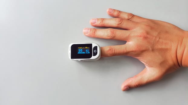
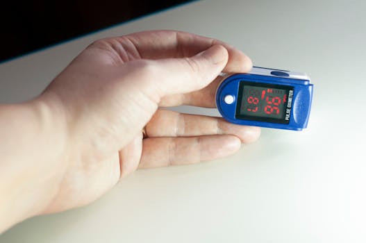
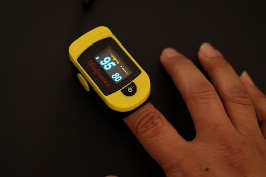
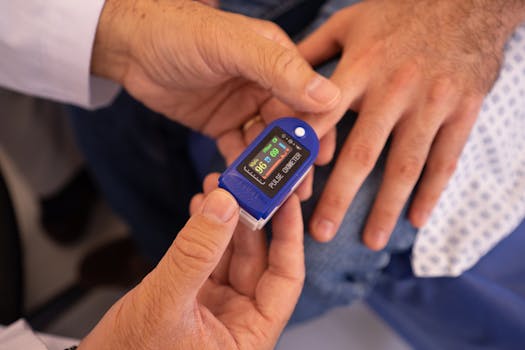
SpO2 (oxygen saturation) is the percentage of hemoglobin molecules carrying oxygen in arterial blood. In most consumer and clinical devices, SpO2 is estimated using pulse oximetry—typically a probe on a finger, ear, or forehead that analyzes light absorption patterns synchronized with pulsatile blood flow.
Because SpO2 is tied to arterial oxygenation, it tends to change when ventilation is insufficient to maintain oxygen levels, when airflow is obstructed, or when gas exchange is impaired. In sleep-disordered breathing, oxygen saturation can drop during apnea or hypopnea events and then recover when breathing resumes.
How pulse oximeters estimate SpO2
Pulse oximeters infer saturation by comparing red and infrared light absorption through tissue. Several factors can distort readings:
- Motion and poor sensor contact (shaking, sleeping position, loose placement)
- Low perfusion (cold extremities, anemia, shock states)
- Skin pigmentation and nail polish (can affect light absorption)
- Ambient light and device limitations
- Arrhythmias and irregular pulse waveforms
These issues can create transient artifacts that look like real desaturation. For interpretation, it is often more reliable to consider patterns and sustained trends rather than single-point dips—unless the dip is large and prolonged or matches symptoms.
Common SpO2 patterns in sleep and respiratory conditions
In obstructive sleep apnea (OSA), SpO2 may show repeating drops synchronized with breathing interruptions. In chronic lung disease, SpO2 may remain lower for long periods, reflecting baseline impaired oxygenation rather than isolated events. In acute respiratory problems, SpO2 can decline more rapidly and may not recover between events.
Two practical points are important:
- Magnitude matters: deeper desaturations generally indicate more severe oxygen transfer impairment during events.
- Recovery matters: fast return to baseline suggests quicker re-ventilation; delayed recovery can indicate ongoing ventilation-perfusion mismatch or persistent obstruction.
ODI: oxygen desaturation index and what it counts
Definition: event counting rather than continuous oxygen level
ODI stands for oxygen desaturation index. It is typically defined as the number of times per hour of sleep (or monitoring time) that oxygen saturation drops by a specified amount from baseline—most commonly ≥3% or ≥4%, depending on the clinical protocol—followed by recovery.
Unlike SpO2, which is a continuous value, ODI is an event frequency metric. It reduces complex oxygen waveforms into a count of desaturation events meeting criteria. That makes ODI useful for estimating how often oxygenation is significantly disrupted, but it also means it can miss certain forms of physiologic stress.
How ODI is derived from oximetry data
While exact calculation details vary by system, ODI generally involves:
- Establishing a baseline saturation (often using a moving reference or pre-event level)
- Detecting desaturation events that meet a threshold (e.g., 3% drop)
- Ensuring the event is discrete (recovery and separation from adjacent events)
- Normalizing to time (events per hour)
This “counting” approach means ODI is sensitive to the threshold chosen. A person may have frequent mild drops that do not cross the threshold and therefore produce a relatively low ODI despite noticeable oxygen variability.
Why ODI often aligns with sleep-disordered breathing
In OSA and related conditions, breathing interruptions lead to reduced ventilation, which can lower arterial oxygen saturation. When these drops are large enough and recur frequently, they produce a higher ODI. Clinicians often use ODI alongside airflow signals, respiratory effort, and symptom history to characterize severity.
However, ODI is not identical to apnea-hypopnea index (AHI). ODI reflects oxygen effects, not respiratory effort patterns directly. Two individuals can have similar AHI values but different ODI values depending on:
- Baseline oxygenation (lower starting SpO2 can change the likelihood of crossing thresholds)
- Duration of events (longer events tend to cause larger desaturations)
- Ventilatory reserve and CO2 sensitivity
- Hemoglobin and circulation factors affecting pulse oximetry accuracy
Limitations of ODI
ODI is powerful, but it has blind spots:
- Oxygen-only view: it does not capture airflow limitation that does not lead to desaturation.
- Threshold dependency: smaller drops may be clinically relevant yet not counted.
- Sensor artifacts: motion or poor signal quality can create false event counts.
- Different protocols: some systems use different desaturation definitions, making cross-device comparisons tricky.
For that reason, ODI should be interpreted as part of a broader monitoring picture—often alongside airflow, respiratory rate, and symptom context.
Respiration rate: timing and pattern of breathing
Definition and why it is not the same as oxygenation
Respiration rate (breaths per minute) measures how frequently a person is breathing. It is fundamentally different from SpO2 and ODI because it reflects ventilation behavior, not the oxygen result of gas exchange.
Respiration rate can increase during anxiety, pain, fever, metabolic acidosis, or hypoxia-driven ventilatory drive. It can also decrease during sleep stages, sedation, neuromuscular weakness, or central breathing disorders. In other words, respiration rate can change for many reasons that do not immediately cause oxygen desaturation.
How respiration rate is measured in monitoring devices
Respiration rate is captured using different sensing methods depending on the device:
- Impedance-based signals (thoracic movement patterns)
- Accelerometer/kinematic detection
- Airflow or pressure sensors (more common in clinical settings)
Each method has different sensitivity to motion, body position, and sensor placement. For example, a wearable that infers breathing from chest movement may misread breathing during significant movement or when the body position changes rapidly.
Respiration rate patterns relevant to sleep and breathing disorders
In sleep-disordered breathing, respiration rate may show:
- Periodic changes around obstructive events (often variable)
- Reduced airflow with compensatory effort that does not always translate into higher rate
- Central breathing patterns where breathing may pause or become irregular, sometimes producing changes in rate
Because respiration rate is a timing metric, it can reveal breathing instability even when SpO2 remains relatively stable—particularly in early disease or in individuals who desaturate less.
SpO2 vs ODI vs respiration rate: how they relate during breathing events
Different “layers” of information
A helpful way to understand SpO2 vs ODI vs respiration rate is to think of them as three layers:
- Respiration rate: how the breathing cycle behaves (ventilation timing)
- SpO2: the oxygen outcome (oxygenation status)
- ODI: how often oxygenation is disrupted enough to meet a threshold (event frequency)
Breathing events can occur without immediate oxygen drops, and oxygen drops can occur without dramatic changes in rate. The relationship is therefore probabilistic rather than deterministic.
Scenario examples (educational, not diagnostic)
Below are common patterns clinicians consider when aligning these signals:
- Frequent breathing disruption with minimal desaturation: respiration rate may show irregularity, but SpO2 stays near baseline and ODI remains low. This can occur when events are shorter, ventilation is partially maintained, or oxygen reserve is adequate.
- Oxygen drops with modest rate changes: SpO2 may fall during obstructive events, yet respiration rate may not clearly spike because breathing can be obstructed without a simple “faster” pattern. ODI can rise if desaturations cross the threshold repeatedly.
- Baseline low SpO2 with variable rate: in chronic respiratory disease, SpO2 may be persistently reduced. ODI may be elevated or less informative depending on how much saturation fluctuates around baseline.
- Artifact-driven changes: motion can produce apparent desaturations that inflate ODI and create misleading SpO2 dips, while respiration rate signals may remain stable (or vice versa).
Why desaturation timing matters
Oxygen saturation does not always drop instantly. During an apnea or hypopnea, oxygen levels fall as oxygen in the lungs and blood is consumed. The speed of that drop depends on event duration and baseline physiology. That is one reason ODI may reflect event duration more than respiration rate does.
In addition, pulse oximeter response time and averaging algorithms can delay or smooth SpO2 changes. As a result, respiration rate may show a breathing interruption earlier than the SpO2 dip becomes visible.
How to interpret trends in real monitoring data
Start with data quality and context
Before interpreting physiology, ensure the signals are trustworthy:
- Check for poor sensor contact, motion periods, or low perfusion events.
- Look at whether desaturation events cluster around movement or device signal changes.
- Confirm that respiration rate values are plausible for the person’s activity state (sleep vs wake vs sitting up).
If a wearable provides signal quality indicators, use them. If not, visually inspect whether SpO2 and rate behave consistently rather than erratically.
Use “direction” and “pattern,” not just single numbers
Single-point readings can be misleading. More informative are:
- Repeated cycling of SpO2 drops and recoveries
- Event clustering (e.g., many desaturations during a particular sleep stage or position)
- Respiration rate instability (frequent irregularities or periodic breathing)
- Trends across the night (worsening toward morning can occur with sleep stage changes or supine position)
ODI is especially useful for comparing monitoring sessions within the same system and protocol, because it summarizes frequency. Still, it should not be treated as a standalone measure of severity without clinical context.
Understand what “normal” means for your situation
“Normal” oxygenation and breathing patterns vary by age, baseline lung function, altitude, comorbidities, and whether monitoring occurs during sleep or wake. For example:
- Some people naturally run lower SpO2 due to chronic lung disease.
- Others may have intermittent desaturations that correlate strongly with sleep position or stage.
- Respiration rate varies by sleep stage and can be lower during deeper sleep.
The best interpretation compares the pattern to the person’s own baseline and to known triggers (position, illness, sedating medications, alcohol use, or nasal congestion).
Practical guidance for improving signal reliability
Optimizing SpO2 and ODI accuracy
Because ODI depends on SpO2 event detection, improving SpO2 signal quality improves ODI reliability. Practical steps include:
- Ensure the probe is firmly placed according to the manufacturer’s guidance.
- Warm the hands or ensure good peripheral circulation if cold exposure is common.
- Avoid excessive motion; consider stable sensor placement and secure straps.
- Remove nail polish if relevant to the sensor type.
If you use a wearable system that supports different sensor modes, choose the mode intended for nighttime monitoring rather than daytime activity.
Optimizing respiration rate measurements
Respiration rate measurements can be influenced by body movement and sensor placement. To improve consistency:
- Wear the device snugly but comfortably to reduce shifting.
- Minimize sensor movement during sleep (adjust straps or positioning).
- Be cautious interpreting respiration rate during periods of significant tossing or sitting up.
When breathing signals disagree with oxygenation signals, consider whether one of the sensors is more affected by motion at that time.
What to do if metrics conflict
Conflicts are common and usually informative rather than confusing:
- Low ODI with irregular respiration rate: oxygenation may remain adequate despite ventilation instability; investigate the breathing pattern with clinical assessment if persistent.
- High ODI with relatively stable respiration rate: oxygen may be dropping during obstructive events that do not produce a simple rate increase; look for cyclic patterns and correlate with symptoms.
- SpO2 dips without consistent event patterns: suspect artifact or transient issues (cold hands, motion) unless dips are large and repeated.
In many cases, clinicians will combine oximetry with airflow or respiratory effort signals to determine whether oxygen desaturation is driven by obstructive, central, or mixed mechanisms.
When to seek clinical evaluation based on monitoring patterns
Monitoring metrics can guide urgency, but they do not replace medical assessment. Consider prompt clinical evaluation if there are signs of significant respiratory compromise, such as:
- Repeated or sustained low SpO2 readings
- Frequent desaturation events (reflected by elevated ODI) especially if accompanied by symptoms
- Marked breathing irregularity with daytime sleepiness, morning headaches, or witnessed apneas
- New symptoms such as shortness of breath, chest pain, confusion, or cyanosis
If SpO2 is very low or symptoms are concerning, follow local emergency guidance immediately. For non-emergent concerns, bring the monitoring report to a clinician so they can interpret it alongside medical history, exam, and—if needed—diagnostic testing such as polysomnography or formal sleep studies.
Prevention and risk reduction: lowering desaturation and supporting ventilation
While metrics can signal problems, prevention focuses on reducing triggers and improving breathing mechanics. Practical steps depend on the underlying cause, but general guidance includes:
- Address nasal obstruction (allergies, congestion) to reduce upper airway resistance during sleep.
- Avoid sedatives and alcohol when they worsen sleep-disordered breathing (clinician guidance is important).
- Sleep positioning: some people experience more events when sleeping supine; positional strategies can reduce episodes.
- Manage chronic respiratory conditions with appropriate medical care and adherence to prescribed therapies.
- Maintain healthy weight when relevant, since excess weight can increase airway collapsibility.
For people using continuous positive airway pressure (CPAP) or other therapies, monitoring metrics are often used to assess response over time. Even then, the most reliable interpretation comes from standardized device reports and clinician review rather than isolated changes in one metric.
Summary: choosing the right metric for the right question
SpO2 vs ODI vs respiration rate reflects three different aspects of breathing and oxygenation. Respiration rate describes breathing timing and pattern. SpO2 shows the oxygenation outcome at a moment in time. ODI counts how frequently oxygen drops meet a threshold, summarizing event frequency over the monitoring period.
When interpreting continuous monitoring data:
- Use respiration rate to understand breathing instability and timing of events.
- Use SpO2 to gauge oxygenation status and recovery behavior, while accounting for sensor artifacts.
- Use ODI to quantify how often desaturation is occurring, recognizing that it depends on the desaturation threshold and event definitions.
Most importantly, look for consistent patterns, correlate metrics with sleep position and symptoms, and seek professional evaluation when monitoring suggests clinically significant breathing or oxygenation impairment.
09.01.2026. 09:51