Autonomic–Immune Link: CRP, IL-6, TNF-α in Stress Physiology
Stress physiology is not just “mental”: it reshapes immune signaling
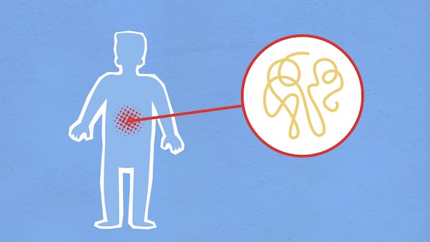
Stress is often described as a psychological state, but biologically it is a coordinated physiological program. When the brain and body detect threat or challenge, the autonomic nervous system (ANS) and the endocrine system change heart rate, vascular tone, energy availability, and—critically—immune activity. This is where the autonomic–immune link becomes central: immune cells do not respond only to pathogens; they also respond to neural inputs and stress hormones.
Three immune-related signals frequently used to track this process are IL-6, TNF-α, and CRP. IL-6 and TNF-α are cytokines involved in inflammation and immune regulation. CRP (C-reactive protein) is an acute-phase protein produced mainly by the liver in response to inflammatory cytokines, especially IL-6. Together, they provide a systems-level snapshot of how stress can shift immune tone.
This article explains the autonomic–immune link in stress physiology using a systems biology lens. You’ll learn the pathways that connect neural control to cytokine production, why CRP rises after inflammatory signaling, and how to interpret these markers in real life. The goal is educational: understanding mechanisms, not diagnosing disease.
The autonomic nervous system as an immune regulator
The ANS has two major branches: the sympathetic system and the parasympathetic system. Both influence immune cells indirectly through neurotransmitters and hormones, and in some contexts directly through innervation of lymphoid organs.
Sympathetic signaling and immune activation
During acute stress, sympathetic output increases. Norepinephrine released from sympathetic nerve terminals can modulate immune cell behavior. Depending on the cell type and receptor profile, sympathetic signaling may enhance inflammatory responses, promote leukocyte trafficking, or alter cytokine production. In practice, this means that persistent or repeated stress can bias the immune system toward a pro-inflammatory pattern in some individuals.
Parasympathetic control and the “cholinergic anti-inflammatory pathway”
The parasympathetic branch—particularly through the vagus nerve—has an inhibitory influence on inflammation. A key mechanistic concept is the cholinergic anti-inflammatory pathway, in which acetylcholine signaling can suppress inflammatory cytokine release in macrophages and other immune cells. This is one reason why measures of vagal tone (and interventions that affect it) are often discussed in the context of inflammation regulation.
From a systems biology standpoint, the ANS functions like a control system: it integrates signals from the brain and body, then applies “set points” to immune activity. When stress is frequent, the control system may spend more time in a state that favors inflammation or less time in a state that favors inhibition.
From neural signals to cytokines: IL-6 and TNF-α as mechanistic nodes

To understand the autonomic–immune link CRP IL-6 TNF-alpha stress physiology relationship, it helps to track the flow of information: neural and endocrine signals influence immune cells; immune cells release cytokines; cytokines drive downstream acute-phase responses.
IL-6: a hub cytokine that links stress to acute-phase biology
IL-6 is produced by many cell types, including immune cells (e.g., monocytes/macrophages) and also non-immune tissues under stress conditions. IL-6 is often described as a “hub” cytokine because it coordinates multiple processes: fever responses, immune cell differentiation, acute-phase protein production, and metabolic reprogramming.
In stress physiology, IL-6 can rise due to a combination of factors: sympathetic activation, glucocorticoid dynamics, tissue stress, and immune activation. IL-6 also acts in feedback loops; it can influence both inflammatory trajectories and how the body resolves inflammation.
TNF-α: an early inflammatory signal with broad downstream effects
TNF-α (tumor necrosis factor-alpha) is another central cytokine. It can be produced rapidly in inflammatory contexts and can amplify immune responses by recruiting and activating additional immune cells. TNF-α is also involved in endothelial activation and changes in vascular function, which affect immune cell movement and tissue-level inflammation.
Because TNF-α can be an early driver, it often contributes to the conditions that later increase acute-phase markers like CRP. However, timing matters: cytokine dynamics are not static, and different cytokines may peak at different times after a stressor or inflammatory trigger.
Why CRP rises: IL-6–driven acute-phase response as a systems readout
CRP is widely measured because it is relatively stable in blood compared with many cytokines and reflects systemic inflammatory activity. CRP is produced primarily by the liver as part of the acute-phase response. The liver’s acute-phase program is influenced by cytokines—especially IL-6.
The acute-phase cascade in stress-linked inflammation
When immune signaling increases (for example, via IL-6 and other inflammatory mediators), hepatocytes shift gene expression toward acute-phase proteins. CRP is one of these proteins. Therefore, CRP can be viewed as a downstream “integrator” of upstream immune signaling.
This integrator role is important in systems biology: CRP may not tell you which cytokine triggered inflammation, but it can indicate that inflammatory signaling has reached a systemic threshold. It also means CRP can rise due to multiple upstream causes, including infection, tissue injury, chronic inflammation, and stress-linked immune activation.
Timing and interpretation: why one measurement may mislead
Cytokines like IL-6 and TNF-α can fluctuate rapidly. CRP rises more slowly and typically declines more gradually. As a result, a single CRP measurement may reflect inflammatory activity from days earlier rather than the immediate state of stress at the moment of blood draw.
For practical interpretation, consider three points:
- Temporal mismatch: cytokines may peak and fall quickly; CRP may lag.
- Non-specificity: CRP increases with many inflammatory processes, not only stress physiology.
- Context matters: recent illness, injuries, sleep disruption, and medication effects can all shift CRP and cytokine levels.
Mechanisms behind the autonomic–immune link: key pathways
The autonomic–immune link is not a single pathway; it is a network of interacting routes. Several mechanisms help connect ANS activity to immune signaling.
Neurotransmitters and immune receptors
Immune cells express receptors for neurotransmitters and hormones. Norepinephrine can modulate immune cell activation states through adrenergic receptors. Acetylcholine and related cholinergic signals can influence cytokine production through nicotinic receptor pathways on immune cells, supporting anti-inflammatory effects in some contexts.
These receptor-mediated interactions provide a direct bridge between neural activity and immune output, making immune cells partially “sensory” to the nervous system.
Vagal pathways and inflammatory reflex concepts
The vagus nerve is often discussed in terms of an “inflammatory reflex,” a control loop that detects inflammatory signals and adjusts neural output to restrain excessive inflammation. While research continues to refine the exact components of this reflex, the overall principle is consistent: neural pathways can detect inflammatory status and modulate immune responses.
Stress hormones as parallel controllers
Although this article emphasizes autonomic pathways, the endocrine system also shapes cytokines. Cortisol and other stress hormones can suppress some inflammatory processes while potentially allowing other pathways to remain active. In chronic stress, altered cortisol rhythms and glucocorticoid resistance in some immune contexts may change the typical anti-inflammatory balance.
Systems biology emphasizes that stress physiology is multi-controller. Neural inputs and endocrine inputs converge on immune cells, and their combined effect determines cytokine output and downstream CRP production.
Systems biology view: feedback loops, thresholds, and heterogeneity

In real organisms, the autonomic–immune link is shaped by feedback loops and individual variability. This is why the same stressor can produce different biomarker patterns across people.
Feedback loops that can amplify or dampen inflammation
Cytokines can influence neural signaling and behavior. For example, inflammatory mediators can affect sleep, appetite, and sickness behavior, which then change stress physiology and autonomic tone. This creates bidirectional coupling: nerves influence immune cells, and immune signals influence brain and autonomic regulation.
Threshold behavior: why “small” changes can matter
Immune systems often operate with thresholds. A mild increase in cytokine signaling might not substantially raise CRP, while a larger or longer-lasting increase may exceed the liver’s acute-phase activation threshold. This helps explain why CRP is sometimes more informative about systemic inflammation than about immediate stress.
Heterogeneity: immune baseline and receptor sensitivity
People differ in baseline inflammation, immune cell composition, receptor expression, and history of exposures. Some individuals may show stronger IL-6 responses to stress, while others show more TNF-α-driven patterns. Some may have more robust cholinergic anti-inflammatory effects; others may have reduced vagal tone or altered receptor responsiveness.
Therefore, interpreting IL-6, TNF-α, and CRP together is often more meaningful than interpreting any single marker alone.
Practical guidance: measuring and interpreting IL-6, TNF-α, and CRP in stress contexts
If you are studying stress physiology or interpreting biomarker results, the practical challenge is separating stress-linked immune changes from unrelated inflammatory causes. The following guidance is educational and general.
Understand what each marker “means”
- IL-6: a cytokine hub that often links upstream immune activation to acute-phase biology; can vary with timing and context.
- TNF-α: an early inflammatory signal that can amplify immune activation; timing can be critical.
- CRP: a systemic acute-phase readout influenced by IL-6 and other mediators; rises later and may persist longer.
Consider timing relative to stress and illness
To interpret stress-linked immune changes, consider when the stressor occurred and whether any infection, injury, dental issues, or inflammatory conditions were present. CRP in particular can remain elevated due to recent illness even if the acute stress has resolved.
Account for confounders that affect inflammation
Common factors that can shift these biomarkers include:
- Sleep disruption and circadian misalignment
- Recent vigorous exercise (which can transiently increase inflammatory signals)
- Body composition and metabolic inflammation
- Smoking and air pollution exposure
- Medications such as corticosteroids or anti-inflammatory drugs
Use patterns rather than single values
For educational interpretation, a trend across time is usually more informative than a one-off measurement. If IL-6 and CRP rise together over a consistent period while TNF-α changes less, that may suggest a particular inflammatory profile. If TNF-α rises early and CRP follows later, that pattern can fit an acute inflammatory trajectory.
However, biomarker patterns should always be interpreted in clinical context by qualified professionals, especially if values are markedly elevated.
How stress physiology can influence inflammation resolution
Inflammation is not only about onset; it is also about resolution. Stress physiology can affect both sides of the equation.
Pro-inflammatory bias under chronic stress
Repeated stress may shift autonomic balance toward sympathetic dominance and reduce effective parasympathetic inhibition. Over time, this can contribute to a pro-inflammatory bias, reflected by higher baseline levels of inflammatory markers in some people.
Impaired recovery: when the “off switch” is delayed
Even after a stressor ends, immune responses may remain elevated longer than expected if neural regulation and endocrine rhythms are disrupted. CRP’s slower kinetics can make this appear as lingering systemic inflammation even when the immediate stress event has passed.
Ways to support autonomic-immune balance through lifestyle physiology
Because the autonomic–immune link is biologically grounded, lifestyle factors that influence autonomic tone and stress physiology can plausibly affect inflammatory signaling. The following guidance is practical and non-prescriptive.
Sleep regularity as a regulator of immune tone
Sleep timing and quality influence cytokine dynamics and autonomic function. Regular sleep schedules help stabilize circadian control, which in turn supports more consistent immune regulation.
Physical activity, but with attention to recovery
Moderate, consistent activity can support autonomic balance and improve metabolic health, both of which influence inflammation. At the same time, excessive training without recovery can temporarily raise inflammatory markers. The key is recovery and gradual progression.
Breathing and vagal tone: what the physiology suggests
Practices that increase parasympathetic activity—such as slow breathing—may influence inflammatory pathways via cholinergic mechanisms. Research findings vary by protocol and population, but the physiological rationale aligns with the concept of vagal anti-inflammatory control.
Stress management that changes physiology, not just perception
Stress reduction strategies can affect bodily stress responses through autonomic and endocrine routes. Techniques that promote downshifting of arousal states—whether mindfulness-based, cognitive-behavioral, or relaxation-focused—may help reduce chronic inflammatory bias in some individuals.
Importantly, these interventions do not “erase” inflammation; they aim to improve the regulation and recovery dynamics of the autonomic–immune system.
When to seek medical evaluation for inflammatory markers
Inflammatory biomarkers like CRP and cytokines can be elevated for many reasons. If you have lab results that are persistently elevated, rapidly rising, or accompanied by symptoms such as fever, unexplained weight loss, persistent pain, shortness of breath, or signs of infection, it is important to seek medical evaluation. A clinician can interpret results alongside medical history, physical examination, and additional tests.
From a systems perspective, elevated CRP does not automatically mean “stress inflammation.” It signals that an acute-phase response is active and that upstream causes should be assessed.
Summary: integrating CRP, IL-6, TNF-α with autonomic control
The autonomic–immune link in stress physiology describes how neural regulation can shift immune activity and inflammatory signaling. Sympathetic and parasympathetic pathways influence immune cells through neurotransmitters and receptor-mediated effects, while endocrine stress hormones provide parallel control. In this network, IL-6 often serves as a key hub cytokine, TNF-α as an early amplifying signal, and CRP as a downstream acute-phase integrator—commonly rising after cytokine-driven systemic inflammation.
Because biomarker kinetics and confounders matter, the most reliable educational approach is to interpret IL-6, TNF-α, and CRP as a coordinated pattern over time rather than as isolated snapshots. Supporting autonomic-immune balance through stable sleep, appropriate recovery, and stress physiology downshifting may help the body regulate inflammatory responses more effectively.
03.05.2026. 12:08